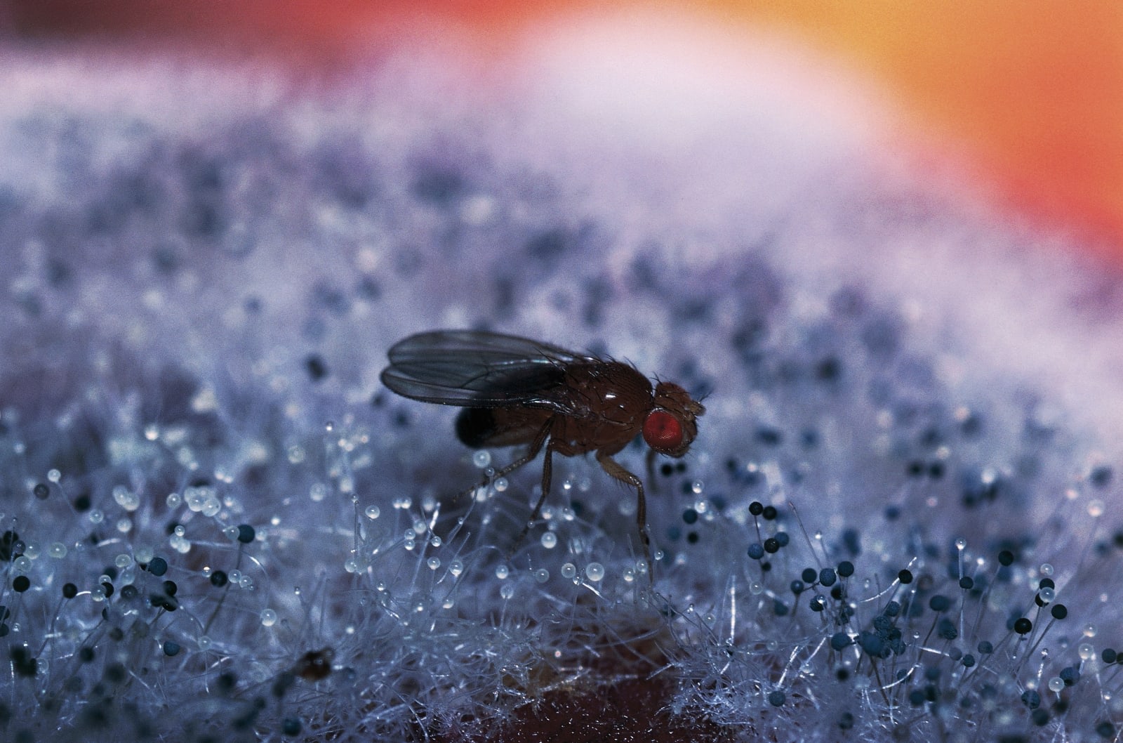
Sure, fully mapping the human brain is impressive, but think about it: our thinking muscle is pretty big. Not to be outdone by this week’s advances from the Allen Institute, scientists from Japan’s Tokai University have made a 3D model of the neurons in a fruit fly’s brain. Think about that for a bit. Exactly; it’s tiny. Okay, ready to read some more? Cool.
As MIT Technology Review tells it, the scientists had to “pickle a fly brain in silver dye, bombard it with x-rays and then measure the way the x-rays are scattered in various directions.” The silver dye is the key here because when it’s attacked with x-rays, it illustrates neural pathways. It’s a process called x-ray tomography.
This, coupled with a computer model that estimates an atom’s position, resulted in a model that displays 100,000 neurons and boasts a resolution of 600 nanometers. All told, it took some 1,700 hours to build.
Making a higher resolution model, according to MIT, would be “prohibitively expensive in terms of human workload,” though. Maybe picking a bigger brain would make things a little bit easier.


